Animating DNA replication
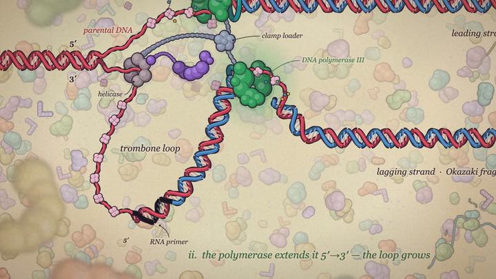
Teaching the central dogma in first-year biochemistry this week, I wanted some lecture animations. An evening with Claude Code got slightly out of hand, and was such a striking example of how one can get to a spectacular output with just a few prompts that I thought I’d share it here.
Tier 1: the cycle
I started with a static slide figure of lagging-strand synthesis: “do you see what’s on this picture?” Claude did.
“Simple and schematic, just the cycle” produced a loop: primer appears, polymerase extends to the previous fragment, exonuclease removes the primer, ligase seals. Repeat.
Tier 2: bells and whistles
“It’s a bit too faithful to the input, add some bells and whistles.” Now there was a replication fork, helicase, base-pair ticks and coloured enzyme blobs working at the appropriate places. The exonuclease ate the primer like Pac-Man. Apparently that was one of the bells.
I asked for a slider between versions. From here, each request became “one notch further on that slider”, which is where the detail tiers came from.
Tier 3: the leading strand
“Also include the leading strand, and make the helicase the ring it actually is.” The picture was becoming a little more complete.
Tier 4: the trombone model
“Next tier, let’s see if you manage: the trombone model.” Here, the lagging-strand polymerase stays at the fork while its template loops out, growing and collapsing like a trombone slide. Claude kept DNA lengths consistent, so older DNA visibly gets reeled in and released.
This took about five feedback rounds, the most of any tier. Fix primase at the fork too, round the kinks, don’t route the DNA through idle enzymes.
Tier 5: prettier
“Make everything slightly more pretty and proper.” Polymerases acquired beta clamps, helicase got six subunits, a clamp loader held the parts together, and primase became cashew-shaped.
Tier 6: go nuts
Tier 5 was what I had hoped to get to. It was correct enough for a first-year bachelor’s class, and to me, the simple schematics made it likely to be immediately intuitive. Mission accomplished. But I had seen Pedro Beltrao post a video of DNA replication animated in 3D by Claude back in September, and the resulting version impressed visually but disappointed a bit on the realism, so I thought I’d give it a quick shot. Since it was 12 at night and I wanted to go to bed, I wrote a bit of a longer prompt, but with the science fixed already to the degree of accuracy that I deemed acceptable, it was focussed purely on the visuals.
“Free rein, go nuts, impress me.” Given about three hours to blow the token budget, Claude launched four parallel sub-agents on different models, reviewed rendered frames and requested several revisions. It took roughly an hour. And boy did it deliver.
Perhaps most spectacular, a cinematic 3D render with double helices and a director’s-cut camera.
Then, most beautiful to me, a painted cell in the style of David Goodsell’s watercolours.
A neon microscope with correctly pairing, glowing base letters.
And a physics sandbox with simulated DNA and adjustable enzyme speeds.
The immediate results required only a few notches to correct some details: detached linkers, SSB emerging from helicase instead of from solution, and a nick obscured by ligase. All fixed.
About 30 prompts, 5,000-plus lines of code I didn’t write, and 1.1 million output tokens later: one static page, with MIT-licensed source. Teaching visuals, accomplished, many times over.
Maximilian JLJ Fürst
Assistant Professor of Computational Protein Design
I research computational protein design and high-throughput protein engineering.